If any of you have ever gone to graduate school, you know it can be stressful and all time consuming; between being a full-time student, a full-time researcher, a part-time teacher (if you’re on a teaching assistantship), while still finding some time to sleep and eat. Maybe on the occasional Friday you find a couple hours to grab a few drinks with your fellow grad students, but other than that grad school is like multiple full-time jobs. Anyway, it was worth the commitment in the end, but when I was in it I needed to find ways to cope with the stress and keep my mental health in check. My way of destressing, while still technically working on my commitments, was to learn Adobe Illustrator (and then open source non-Adobe systems) and create my own figures for presentations and papers. So, here are some figures I created, starting in school, and what they were created for.
As an undergraduate student I worked in a lab that conducted brine shrimp research. For my first peer-reviewed publication I taught myself how to use Adobe Illustrator so I could create figures of all the abnormal stages of development. After those serious figures were done, I had a little fun with the figures and put a twist on my love for the Teenage Mutant Ninja Turtles. I give you: The Juvenile (teenage) Abnormal (mutant) Brine Shrimp.
Brittle Stars
During my first two years of grad school I studied the affects of acidification on aquatic animals. Including brittle stars. Brittle stars are like sea stars but with thinner “brittle” arms. They can reproduce asexually (by themselves, all on their own) by splitting themselves in half. This diagram is showing the first brittle star in blue. It would have been exposed to ocean acidification then removed from it. It is then cut in half and and the missing half grows, in orange. They are then split in half again and the all orange ones would be all new growth that was never exposed to ocean acidification. I would then test them to see if they were affected by their parental exposure.
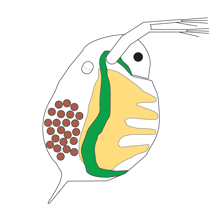
Water Flea (Daphina sp.)
This was also part of my work on acidification on the offspring of asexually reproducing animals. This is the water flea which is freshwater so it would be exposed to lake acidification instead of ocean acidification.
When water fleas are in their eggs, they already have babies in their own eggs. This is showing three generations. The adult water flea is the first generation. The brown circles are the eggs which is the second generation, and inside each egg is a water flea carrying more eggs which are the third generation.
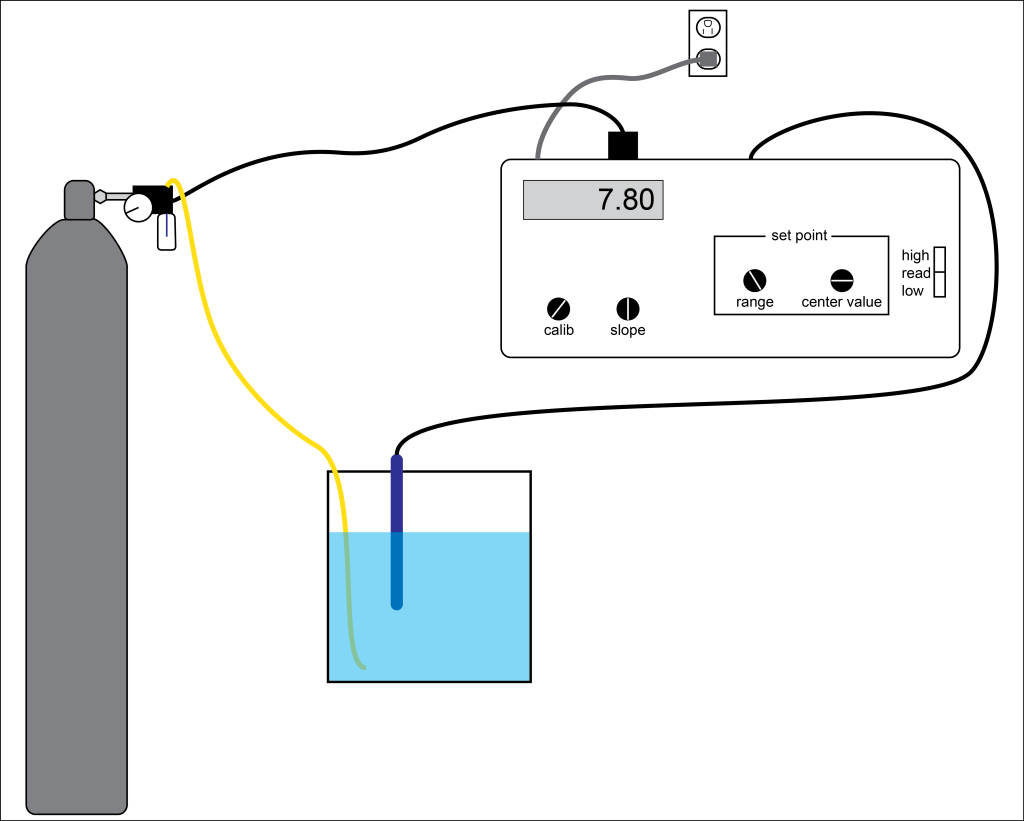
This is showing my water flea set up for how I made the water acidic and how I controlled it. The big gray cylinder contains Carbon Dioxide gas. The rectangular monitor shows the pH (acidification) level. It knows the pH from the blue pH probe in the water. When the pH gets too high the monitor will open up a valve on the cylinder and carbon dioxide will slowly flow into the water through the yellow tube until the pH monitor reads the correct pH in the water again. Then the valve on the cylinder will close and carbon dioxide will stop entering the water.
Mouse (Mus musculus)
In a genetic class in graduate school I had to create a mock research project and present the proposal. Our project used mice so I created this cute, angry looking, mouse.
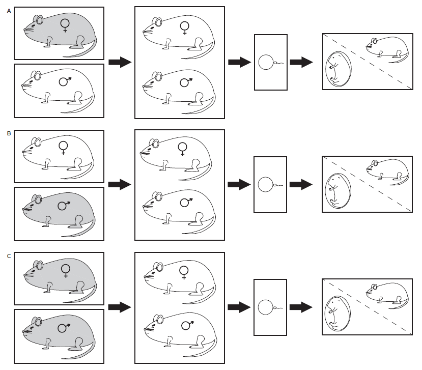
The actual diagram from that mock proposal for that genetics class. Hypothetically mice (in gray down the first column) would be exposed to nicotine. The white mice in the first column were not exposed to nicotine. In group (A) only the females were exposed, in group (B) only the males were exposed, and in group (C) males and females were exposed. All exposed mice were then removed from the nicotine exposure. After a set time after being removed they were allowed to reproduce. We then compare the offspring of each group to see the effects of parental nicotine exposure.
Amphibians
I believe these were completed for a mock research project in a graduate class too. Tadpoles (the above diagram) mainly breath through gills in the water. Frogs (bottom diagram) mainly breath through lungs. All amphibians secondarily breath through their skin. Late stage tadpoles can also gulp air as their lungs develop. I believe this diagram was created to show how we would test the amount of oxygen consumed by tadpoles and by frogs. This is one of my only diagrams I don’t remember all the details of.
Salamander and frog metamorphosis is slightly different. They both start out as gelatinous eggs. Salamander larvae retain external gills all the way through metamorphosis, whereas frog tadpoles quickly develop internal gills after hatching from their eggs. Salamanders develop their front limbs first, and then their back limbs. Frog develop their front and back limbs at the same time, however their front limbs develop under their skin so it looks like their back limb develop first. As salamanders develop their back limbs, their external gills dissolve. As frogs front limbs emerge their tail dissolves. Eventually the salamanders and frogs have completed metamorphosis.
This was drawn for my Masters thesis. It shows the difference between lungs in a frog and a gas bladder in a fish. The first difference, there are two (paired) lungs, whereas there is one fish gas bladder. The second main difference, the frog lungs develop below the pharynx, whereas the fish gas bladder is above the pharynx.
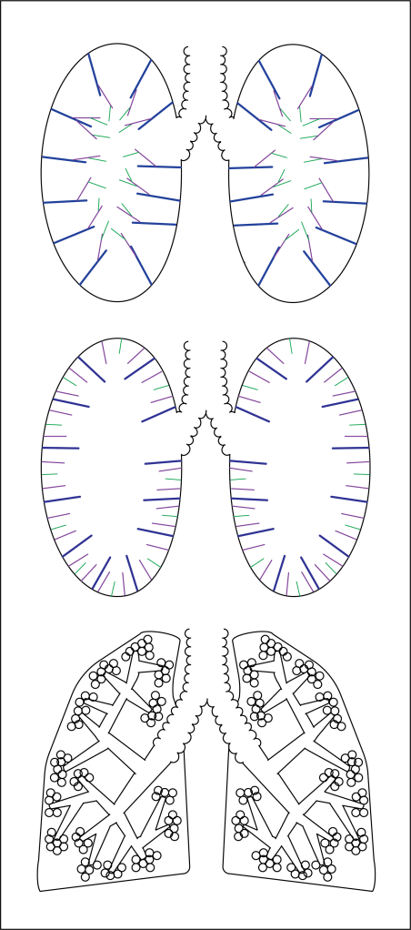
This was also completed for my Masters thesis. These show lung anatomy. The top one is the first hypothesis for internal amphibian lung anatomy. The middle one is the second hypothesis for internal amphibian lung anatomy. The bottom one is the internal anatomy of mammal lungs, like us humans (assuming you are a human reading this and not a frog).
Others
This dancing daisy was originally drawn specifically for a previous article on this website about plant movement. Although the one in the article features different colors on the flower, this one is the original. Check out the article here!
This frog could probably be included in the frog section, but I wanted to put it last as this is the logo created first for my YouTube Channel and then used for this website. The original frog was created for the respirometry drawings in the frog section and then used with slight edits for basically every other frog drawing after that.
Citations
Neumeyer, C.H., J.L. Gerlach, K.M. Ruggiero, and J.A. Covi (2015). A Novel Model of Early Development in the Brine Shrimp, Artemia franciscana, and Its Use in Assessing the Effects of Environmental Variables on Development, Emergence, and Hatching. Journal of Morphology 276:342-360.
Neumeyer, C.H. (2018). Investigating Interspecific and Intraspecific Variation in Lung Development in Amphibians [Master’s Thesis, James Madison University] JMU Scholarly Commons.
AKA: Courtney The Frogologist. AKA: Courtney Neumeyer.
Courtney started this site to provide free science/nature education to all. After taking a break from school, Courtney received her B.S. degree in Dec 2013 from the University of Wisconsin Stevens Point (UWSP). She had a double major in (1) Biology and (2) Wildlife Ecology: Research and Management. Courtney then received her M.S. degree in May 2018 from James Madison University (JMU). Her research thesis investigated the lung development in amphibian eggs, larvae, metamorphs, and adults. Courtney is a co-author on four peer-reviewed scientific research articles under the name Courtney H. Neumeyer. Since grad school Courtney has worked as an environmental educator, conservation educator, recruiter, technical writer, and STEM educator. Courtney has also lived all over the USA.
View more posts